Crohn's Disease Symptoms and Diagnosis Background
Crohn's Disease

Crohn's Disease Symptoms & Diagnosis

An accurate diagnosis is the first step toward proper treatment.

Definition & Statistics

A chronic inflammatory bowel disease
that affects all layers of the digestive tract

Crohn's disease is an autoimmune condition and, along with ulcerative colitis, is a type of Inflammatory Bowel Disease (IBD). Crohn's disease is an inflammatory bowel disease where inflammation occurs throughout the entire digestive tract, from the mouth to the anus, and affects all layers of the bowel wall. It primarily occurs in the duodenum, small intestine, and large intestine, and clinical symptoms vary depending on the site of inflammation.

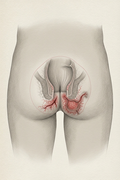
Unlike ulcerative colitis, where inflammation is limited to the mucosa and submucosa, in Crohn's disease, inflammation can occur across all layers of the bowel wall, which can lead to wall edema, strictures, obstruction, perforation, fistulas, and anal fissures. Inflammation occurs discontinuously and can also manifest symptoms outside the gastrointestinal tract, such as in the skin and joints.

About 20-30% of cases occur in childhood and adolescence, with the highest incidence between the ages of 15 and 35. Onset during childhood and adolescence can impair growth. Additionally, Crohn's disease is characterized by poor wound healing in cases of fistulas or anal fissures, including at surgical sites.

Crohn's Disease Surgery Statistics

* 80% experience bowel resection within 20 years of diagnosis
* 20~40% of patients undergo their first surgery within 3 years
* 28% of surgical patients require reoperation within 5 years, with 68% of those requiring it within 2 years

Etiology

Causes involve complex interactions of various factors

It is known to be caused by a complex combination of genetic, immunological, and environmental factors.

GENETICS

Genetic Predisposition

Genetic mutations such as IL23R and
the influence of family history

IMMUNITY

Immune Dysfunction

Overactivated immune response (Th1/Th17, etc.),
and increase in inflammatory cytokines (TNF-α, IL-12/23, etc.)

MICROBIOME

Microbiome Changes

Gut microbiota imbalance and
reduction in beneficial gut bacteria

ENVIRONMENT

Environmental Factors

Smoking, diet, stress,
antibiotic exposure, etc.

Symptoms

Major Symptoms

Depending on the location of the inflammation, various symptoms and diagnostic tests are required.

Gastrointestinal Symptoms

Gastrointestinal Symptoms

Chronic diarrhea, abdominal pain, weight loss, and bloating occur.

Abdominal pain and vomiting due to intestinal strictures or obstruction may occur. Chronic inflammation of the bowel wall can lead to fibrosis, resulting in narrowing (stricture) or blockage (obstruction) of the tract. In modern Western medicine, it is generally considered that strictures or obstructions caused by advanced fibrosis are irreversible and cannot be fully recovered.
Perianal Lesions

Perianal Lesions

- Perianal fistula
- Anal abscess
- Anal fissure
Systemic Symptoms

Systemic Symptoms

- Fatigue
- Fever
- Loss of appetite
- Growth retardation/reduction
Extra-intestinal Symptoms

Extra-intestinal Symptoms

- Arthritis
- Dermatitis (Erythema nodosum)
- Ocular inflammation (Uveitis)
- Hepatobiliary disease

"Because Crohn's disease can cause inflammation throughout the digestive tract and across all layers of the bowel wall,
various diagnostic methods are necessary."

Diagnosis

Types of Diagnostic Tests

Because Crohn's disease can cause inflammation throughout the entire digestive tract and across all its layers, unlike ulcerative colitis, multiple diagnostic methods are required.

1. Clinical Symptoms

Check for diarrhea, abdominal pain, weight loss, perianal lesions, etc.

2. Endoscopy

  • Colonoscopy: Checks for suspected inflammation in the large intestine.
  • Gastroscopy: Checks for suspected inflammation in the stomach and duodenum.
  • Capsule Endoscopy: Checks for suspected inflammation or bleeding in the small intestine.

* Identifies discontinuous inflammatory lesions, longitudinal ulcers, cobblestone appearance, strictures, and fistula openings. Endoscopy can only confirm inflammation on the inner lining of the intestinal wall.

3. Stool Test

Measures fecal calprotectin (an indicator of intestinal inflammation) and performs a fecal occult blood test. If inflammation occurs in the duodenum or small intestine, fecal calprotectin levels may appear low.

4. Biopsy

Analyzes changes in mucosal tissue collected during endoscopy.

5. Blood Test

Checks for elevated inflammatory markers like CRP and ESR, and for anemia. However, since blood inflammatory markers are often normal even when intestinal inflammation is present, its reliability is low.

6. Imaging Tests

CT, MRI, or contrast-enhanced imaging is essential when intestinal obstruction or strictures are present. Fistulas and abscesses can be diagnosed and evaluated via CT, MRI, or ultrasound.

Risks and Limitations to Consider During Testing

Risks of Endoscopy

1. Disruption of Gut Flora: Bowel preparation agents taken before a colonoscopy have an effect similar to washing out the entire normal microbiota, which can destroy gut bacteria. It can take months for the microbiome to recover.

2. Risk of Bowel Damage: Depending on the operator's skill, the endoscope can scrape or bump the bowel wall during insertion, causing injury or, in severe cases, intestinal perforation.

3. Limitations of Capsule Endoscopy: There are cases where a swallowed capsule endoscope gets stuck in the digestive tract and does not pass through.

Risks of Imaging Tests

Radiation Exposure: Excessive radiation exposure can be a problem with repeated CT scans.

Contrast Toxicity: Contrast agents used to evaluate intestinal strictures can have renal toxicity.

Key Diagnosis

Fecal Calprotectin Test
(FC)

A highly reliable biological indicator with
over 80% correlation with colonoscopy

Fecal calprotectin is a biological indicator secreted by neutrophils, a type of white blood cell involved in intestinal inflammation. Calprotectin is secreted when there is inflammation in the gut and is absent when there is no inflammation. Because the amount secreted varies with the degree of inflammation, it is widely used for the diagnosis and follow-up of inflammatory bowel disease.

Various studies show it to be a reliable marker with approximately 93% sensitivity and 96% specificity. In particular, it shows high reliability, with over 80% correlation with colonoscopy findings (i.e., if calprotectin levels are bad, colonoscopy results are generally bad, and if calprotectin levels are good, colonoscopy results are generally good).

* Note: If there is no inflammation in the colon but only in the duodenum or small intestine, or if there is inflammatory edema on the intestinal wall, the reliability or diagnostic value of calprotectin may decrease. Levels can also rise in cases of bacterial/viral gastroenteritis, celiac disease, lymphoma, or food allergies, so differential diagnosis is necessary.

Reference Guide (mg/kg)
Normal 50.0 ~ 100.0 or below
Borderline 100.0 ≤ Measured Value ≤ 200.0 ~ 250.0
* Checking for differential diseases is essential
Inflammation 200.0 ~ 250.0 or above

Limitations of Blood Inflammatory Markers

Blood inflammatory markers have low reliability. In clinical practice, inflammatory substances on the surface of the intestinal mucosa rarely penetrate the bowel wall to affect the blood in a way that manifests in tests, so blood markers often appear normal even when inflammation is severe on endoscopy. However, if there is wall inflammation, fistulas, or abscesses, they may directly affect the blood, causing CRP and ESR markers to rise.

Levels rise in infectious diseases, but since ulcerative colitis and Crohn's disease are not infectious, they are often normal. Long-term, high-dose use of immunosuppressants can cause leukocyte counts to drop, leading to reduced immunity. Levels may rise if there is an anal fistula or abscess in Crohn's disease.

An acute-phase reactant synthesized and secreted into the bloodstream within hours of the onset of infection or inflammation. It can also rise due to heart attack, sepsis, viral infection, or intense exercise. It can check the level of inflammation in the acute phase. Generally, the normal range is < 0.5 mg/dL, but reference values should always be checked as units may differ.

When blood is drawn and allowed to settle, red blood cells accumulate at the bottom and separate from plasma. At this time, changes in the electrical charge on the surface of the red blood cells due to inflammation levels affect the settling speed. The measured value of this speed is ESR. The average normal value is <20mm/h, which varies by age and gender, and reflects long-term inflammation.

Major Complications of Crohn's Disease

  • Intestinal Stricture
  • Intestinal Perforation
  • Entero-enteric or Enterocutaneous Fistula
  • Intra-abdominal Abscess
  • Nutritional deficiency and growth failure due to malabsorption

F.A.Q

Frequently Asked Questions

Loading FAQ data...
Inquiry AI